Location(s): Holton, Onaga, St. Marys, Frankfort
Whether you or a loved one needs a mammogram, ultrasound, x-ray, CT, MRI or other diagnostic imaging procedure, you want accurate results. At Community HealthCare System, our skilled technicians use the latest high-resolution imaging technology and techniques to provide clear, precise and detailed images for better diagnosis and improved treatment. Our compassionate, professional staff takes great care to provide for your comfort while diagnosing your condition as quickly, conveniently and safely as possible.
For your convenience, CHCS also uses a web-based picture archive and communications system (PACS) that enables us to deliver timely access to high-quality images and interpretations anytime, from anywhere.
Imaging Technologies and Services
Community HealthCare System diagnostic imaging uses state-of-the-art imaging technology and techniques to capture images with a level of detail, clarity and speed never before possible. These ultimately help our technicians to quickly and accurately diagnose your condition.
Computed Tomography (CT)
The CT scanner is an x-ray camera that very quickly takes multiple cross-sectional images inside of the human body. It then carefully reconstructs the images to obtain multiplane pictures.
Featuring one of the most sophisticated multi-slice CT technologies available, CHCS’s Aquilion Prime 40-slice scanner can capture precise images of any area of the body in as little as a fourteen second breath-hold. The fast scanning capabilities and unmatched image quality offer significant benefits for a quick and accurate diagnosis of trauma patients experiencing chest pain or stroke. With this scanner, images can be obtained in seconds, and the images allow physicians to see internal injuries and disease in greater detail than ever before.
X-ray
Radiography, or as it is most commonly known, an x-ray, is the oldest and most frequently used form of medical imaging. An x-ray takes a picture of the bones inside the human body. Each picture takes just a couple of seconds, like a regular digital photograph.
X-ray imaging is the fastest and easiest way for a physician to view and assess broken bones, joint problems or spine injuries. At least two images (from different angles) are taken and often three images are needed if the problem is around a knee, elbow or wrist. X-rays also play a key role in guiding orthopedic surgery and in the treatment of sports-related injuries. X-ray may uncover more advanced forms of cancer in bones, although early screening for cancer requires other methods.
Mammography
Mammography uses special x-ray machines to find tumors in breast tissue. Mammograms are a vitally important tool in early detection of breast cancer. State-of-the-art 3D digital mammography takes only a few minutes, and it can detect tumors before a patient would feel a lump or notice symptoms. Mammograms are read by board-certified radiologists using state-of-the-art monitors. When breast cancer is detected early (stage 1), it’s highly treatable.
Ultrasound
Ultrasound imaging, also called ultrasound scanning or sonography, is a method of obtaining images from inside the human body through the use of high-frequency sound waves. The reflected sound wave echoes are recorded and displayed as a real-time visual image. No ionizing radiation (x-ray) is involved in ultrasound imaging. Obstetric ultrasound refers to the specialized use of sound waves to visualize and thus determine the condition of a pregnant woman and her child.
Ultrasound is a useful way of examining many of the body's internal organs, including (but not limited to) the heart, liver, gallbladder, spleen, pancreas, kidneys and bladder. Because ultrasound images are captured in real time, they can show movement of internal tissues and organs and enable physicians to see blood flow and heart valve functions. This can help to diagnose a variety of heart conditions and to assess damage after a heart attack or other illness.
Fluoroscopy
Fluoroscopy is a technique for continuous or intermittent x-ray monitoring—it’s like a moving x-ray. X-ray images may be viewed directly without taking and developing x-ray photographs. This allows observation of certain dynamic body processes and is useful in certain surgical and diagnostic procedures.
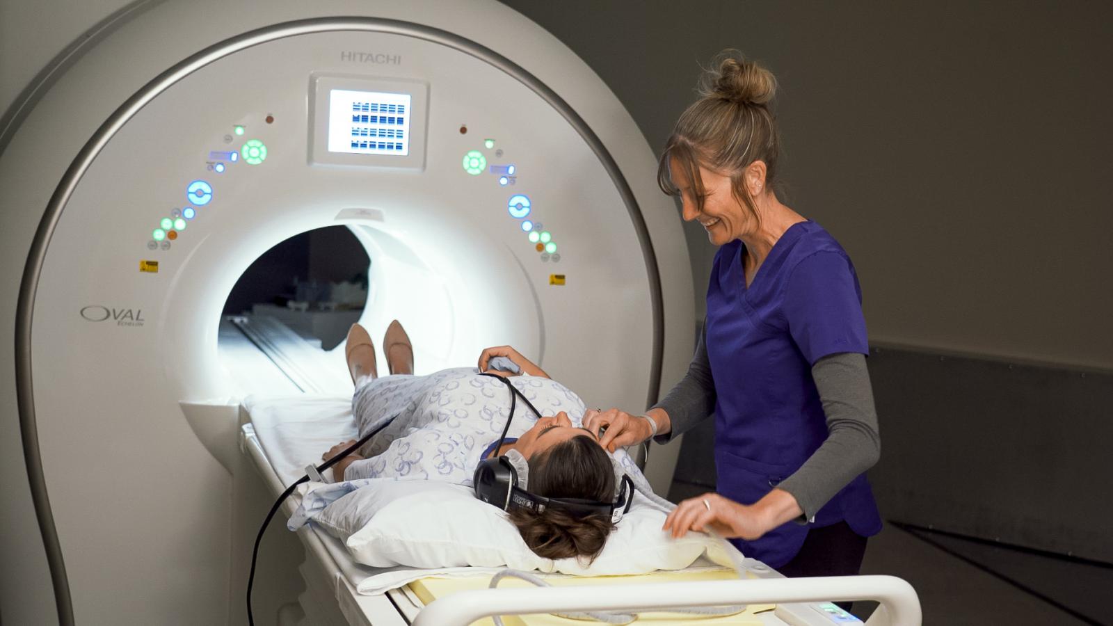
Magnetic Resonance Imaging (MRI)
The MRI machine uses magnets, radio waves and a computer to take very detailed pictures of the inside of the human body. MRIs are useful in finding problems such as tumors, bleeding, injury, blood vessel diseases or infection. MRI also may be done to provide more information about a problem seen on an x-ray, ultrasound scan or CT scan. Contrast material may be used during MRI to show abnormal tissue more clearly. Our MRI offers the widest opening on the market and other features that make it a comfortable experience for our patients. Learn more.
Nuclear Medicine
Nuclear medicine is a subspecialty within the field of radiology. It comprises diagnostic examinations that result in images of body anatomy and function. The images are developed based on the detection of energy emitted from a radioactive substance given to the patient, either intravenously or by mouth. Generally, radiation to the patient is similar to that resulting from standard x-ray examinations.
Nuclear medicine images can assist the physician in diagnosing diseases. Tumors, infection and other disorders can be detected by evaluating organ function.
Contrast
Some procedures may require you to ingest a contrast material. Contrast is a fluid that you will drink or get through an IV (or both). Once it is inside your body, it looks extra bright in the radiology images and makes it easier for the physicians to see what they’re looking for and more accurately diagnose the problem.
Whatever your diagnostic imaging need, CHCS is here for you.
Contact Us
Community HealthCare System
Imaging Services
Onaga: 785-889-4657, ext. 2110
St. Marys: 785-437-3407, ext. 1724
Holton: 785-364-3205, ext. 1444
For Scheduling Services, simply contact:
Patient Access Services
785-889-4272, ext. 2102 or 1-800-889-4274